Stem cells have two key characteristics. They are able to self-renew (proliferate without differentiating or aging) and they can differentiate into mature cells of different lineages – hence the byline “Divide and Differentiate” in the title of this blog. These two properties allow stem cells to repair and regenerate tissues and organs. Stem cells thus form the corner-stone of tissue engineering. The underlying biological mechanisms that regulate stem cell function and self-renewal are fairly complex and our knowledge of these processes remains very limited. The field of stem cell research has generated a lot of enthusiasm, but this enthusiasm can at times skew the interpretation of the actual scientific findings. The hope of being able to regenerate or rejuvenate tissues taps into the old human quest for immortality. This may be the reason why we often come across reports and discussions suggesting that stem cell therapies have a miracle-like quality and will soon be able to cure most diseases. While the ground-breaking discoveries in stem cell biology are indeed fascinating, it is also important to have a realistic view of stem cell therapies and realize that much of stem cell biology is still in its infancy. Premature attempts to use stem cells for clinical therapies are probably not going to succeed.
“The Next Regeneration” started out as part of the Scilogs blogging network and focused on novel developments in stem cell research, regenerative biology and regenerative medicine. Over the years, the topics discussed were expanded to include various aspects of cell biology, psychology, philosophy as well as broader questions related to the process of scientific discovery and scientific communication. It is now an independent blog hosted on this website.
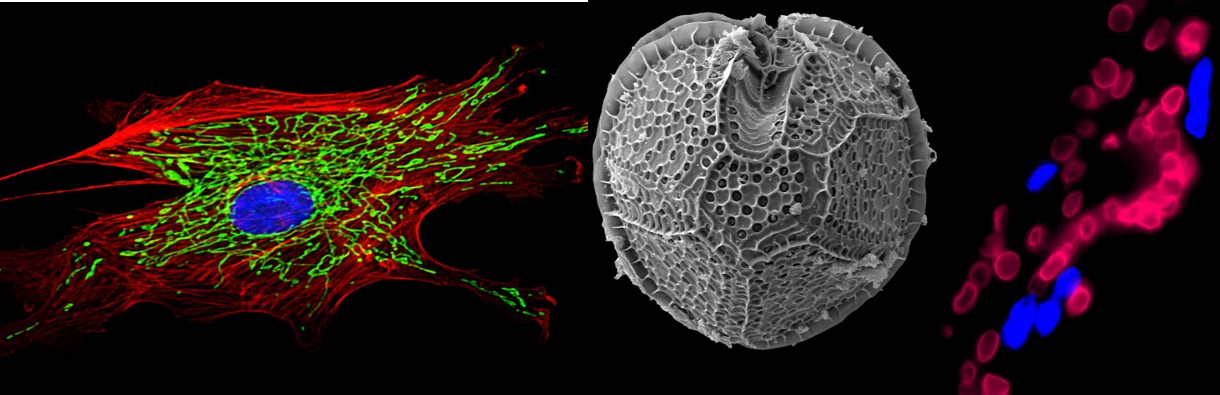
The header image symbolizes three representative projects that we have worked on: On the very left, you can see a fluorescence microscopy image of blood vessel cell in which the mitochondrial network is labeled with green fluorescence; the center shows an electron microscopy image of the algae Gonyaulax polyedra (which has been renamed to Lingulodinium polyedrum) that was used in many of the landmark studies on how biological clocks work; the right image of the header is a confocal microscopy image of a human blood vessel we engineered and implanted into a mouse. The red blood cells are mouse cells flowing through this engineered blood vessel.
